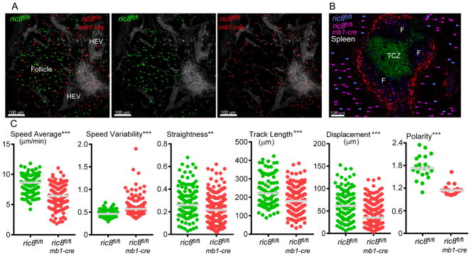
Figure 5.

Intravital microscopy of the inguinal lymph node and imaging of spleen sections reveal that loss of Ric-8A leads to B cells with decreased motility that access B cell niches poorly. (A) Images acquired during intravital microscopy of the inguinal lymph node of a recipient mouse that had received an intravenous transfer WT (green) and ric8Afl/flmb1-cre (red) B cells (1:1 ratio) 18h prior to imaging. Evans blue was infused intravenously immediately before the imaging to outline blood vessels (grey). (B) Standard confocal microscopy of a thick spleen section from the same mouse used for the intravital microscopy focused on a splenic primary follicle. The T cell zone and B cell zones are indicated. The section was immunostained for CD4 (green) and CD169 (red). ric8fl/flmb1-cre B cells (pink) and ric8fl/fl (blue) B cells are shown. The location of the transferred B cells in the surrounding red pulp is indicated with arrowheads of the corresponding color. (C) Motility parameters of the ric8fl/flmb1-cre B cells and ric8fl/fl (control) B cells in the lymph node follicle of the recipient mice. Also shown are polarity measurements (long axis/short axis) of the control and ric-8fl/flmb1-cre B cells. Results are based on the analysis of two separate imaging experiments. ** p < 0.005; *** p < 0.0005.
