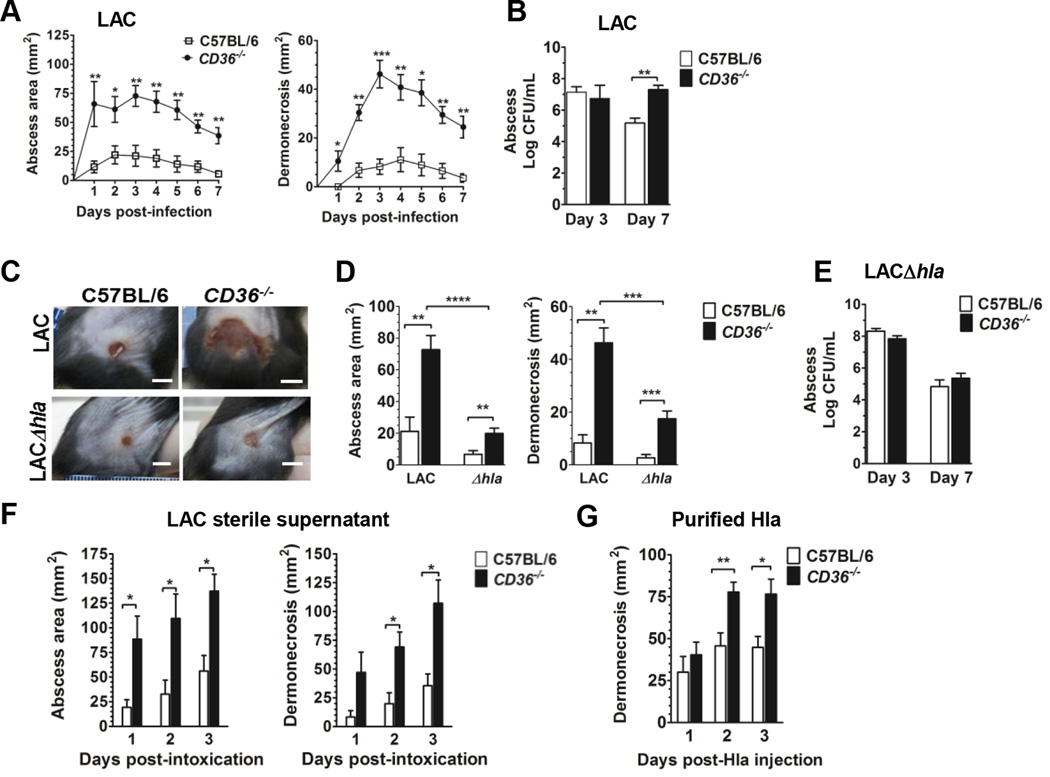
FIGURE 1.
CD36−/− mice have increased dermonecrosis in response to S. aureus infection and alpha hemolysin. (a) Area of abscess (left), dermonecrosis (right) and (b) bacterial burden of mice subcutaneously infected with LAC (8 × 10⁶ CFU). N=8 mice per group from two independent experiments. (c) Representative images of infection sites taken on day 3 post-infection (LAC, 8 × 10⁶ CFU; LACΔhla 1.1 × 10⁷ CFU) (scale bar = 5 mm). (D) Abscess area and dermonecrosis on day 3 post-infection with LAC or LACΔhla. N=8 (LAC) and 20 (LACΔhla) mice per group from two and four independent experiments, respectively. (E) Bacterial burden at the site of infection 3 and 7 days post-infection with LACΔhla (1.1 × 10⁷ CFU). N=8 mice per group from two independent experiments. (F) Mice were intoxicated by subcutaneous injection with LAC sterile supernatant (18 hour culture). Area of abscess and dermonecrosis were measured daily. N=6 mice per group. (G) Area of dermonecrosis of mice injected subcutaneously with 1 µg of purified Hla. N=8 mice per group from two independent experiments. Data shown as mean + SEM. *, p<0.05; **, p<0.01; ***, p<0.001; ****, p<0.0001.

