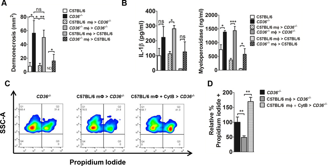
FIGURE 4.
CD36+/+ macrophages negatively regulate the dermonecrotic phenotype of LAC intoxicated CD36−/− mice. Mice were intoxicated by subcutaneous injection with LAC sterile supernatant. 4 h later, mice were either left untreated or peritoneal macrophages were injected adjacent to the site of intoxication. (A) Area of dermonecrosis and local (B) IL-1β and MPO levels measured day 1 post-intoxication. Data reported as mean± SEM. N=4–6 mice per group from two independent experiments. (C,D) Membrane integrity of Ly6G+ cells from intoxication site skin sections measured by flow cytometry as percent propidium iodide uptake relative to CD36−/− controls. N=3–4 mice/group. ns, not significant; ND, not detected; *, p<0.05; **, p<0.01; ***, p<0.001.

