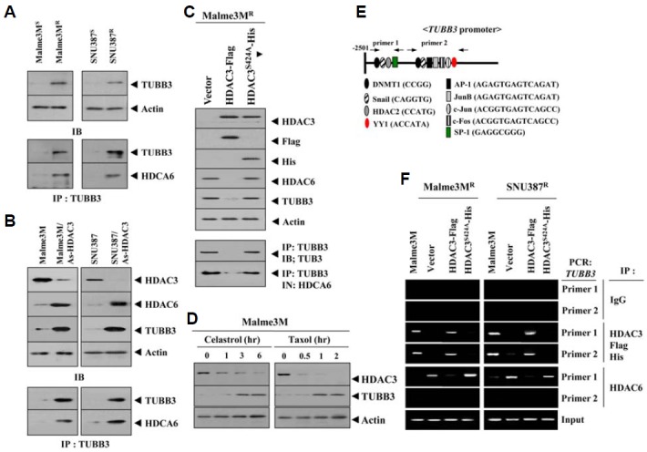
Fig. 4.

HDAC3 regulates expression of tubulin β3 and the interaction between HDAC6 and tubulin β3. (A) Cell lysates from each cell line were immunoprecipitated with the indicated antibody (2 μg/ml), followed by Western blot (lower panel). Cell lysates were also subjected to Western blot (upper panel). (B) Cell lysates of the indicated cell line were immunoprecipitated with the indicated antibody (2 μg/ml), followed by Western blot (lower panel). Cell lysates were also subjected to Western blot (upper panel). (C) At 48 h after transfection with the indicated construct, cell lysates were immunoprecipitated with the indicated antibody (2 μg/ml), followed by Western blot (lower panel). Cell lysate were also subjected to Western blot (upper panel). (D) Malme3M cells were treated with celastrol (1 μM) or taxol (1 μM) for various time intervals. Cell lysates prepared at each time point were subjected to Western blot analysis. (E) Shows the proximal promoter sequences of tubulin β3. (F) At 48 h after transfection with the indicated construct, ChIP assays were performed. Cell lysates prepared from untransfected Malme3M or SNU387 cells were also subjected to ChIP assays.
