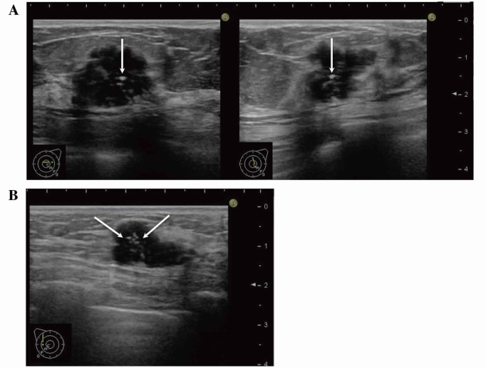
Fig. 2.
Microcalcifications on US images.
A: Transverse (left) and longitudinal (right) US images of the AC area of the left breast. Noncontinuous echogenic spots (arrows) were observed on orthogonal tomographic images.
B: Longitudinal image of the C area of the left breast. Punctuate echogenic spots (arrows) with a tissue composition clearly different from that of the mammary glands were observed inside the mass.
US, ultrasonography.

