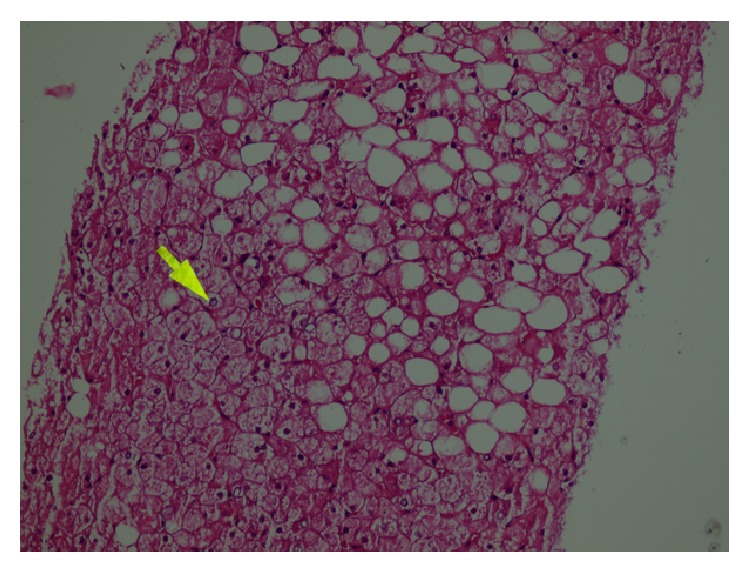
Figure 1.

Histological findings in the liver (hematoxylin and eosin stain). The hepatocytes are diffusely swollen with rarefaction of cytoplasm and accentuation of the cell membranes. Numerous hepatocytes exhibit glycogenated nuclei. There are fatty droplets presenting without inflammation and fibrotic changes.
