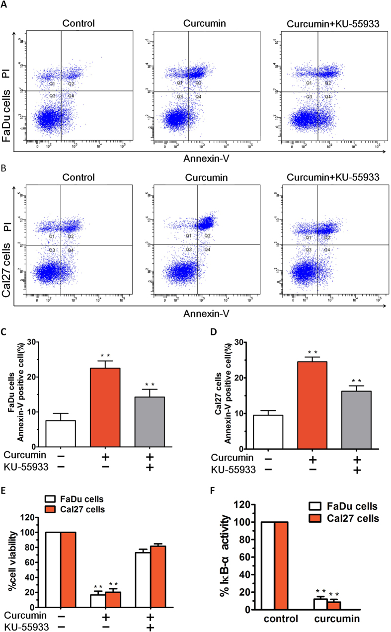
Figure 6. Annexin-V/PI staining showed that curcumin treatment induced apparent early and late apoptosis in HNSCCs.
(A,B) After treatment with curcumin (7 μM), the FaDu cells and Cal27 cells groups showed early and late apoptosis at rates of 17.20% and 21.60%, respectively. (C,D) After treatment with 10 μM KU55933, the apoptosis rate was significantly reduced, not only in FaDu cells group, but also in Cal27 cells group. (E) FaDu and Cal27 cells were treated with curcumin in the presence or absence of the ATM inhibitor KU55933 for 24 h, followed by assessment for cell viability using MTT assays. (F) FaDu and Cal27 cells were treated with curcumin (7 μM) for 24 h, and extracts were then analysed for NF-κB activity by measuring phosphorylated IκB-α. Data presented are means ± SD (n = 3; P < 0.05).

