Figure 1.

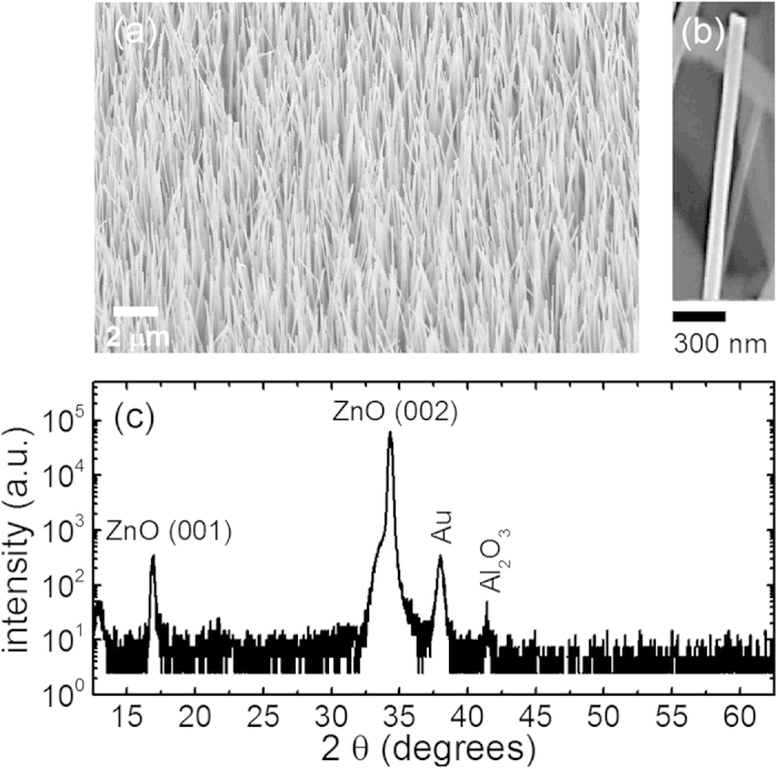
(a) An SEM overview image of the studied ZnO NWs under a tilted view of 45°. (b) a magnified SEM image of a single ZnO NW. (c) XRD diffractogram of the studied ZnO NWs.

(a) An SEM overview image of the studied ZnO NWs under a tilted view of 45°. (b) a magnified SEM image of a single ZnO NW. (c) XRD diffractogram of the studied ZnO NWs.