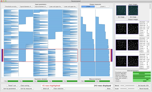
Figure 5.

The results of a nuclei detection algorithm on photomicrographs of human HT29 cells (colon cancer). The data have been sorted on the sixth column, which encodes the number of cells detected in the first input image. The highlighted rows indicate results where nuclei detection is correct and have been validated by also considering the output images at the far right. This reveals relationships with the values taken for the second and fourth input parameters (column two and four).
