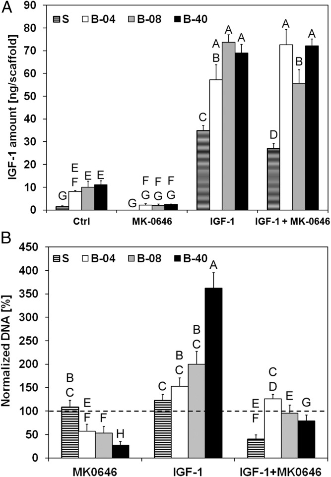
Fig. 4.
Effect of flow-derived shear stress on ES cell drug sensitivity. IGF1 secretion and drug sensitivity for experimental groups tested at different flow rates (S, static; B-04, 0.04 mL/min; B-08, 0.08 mL/min; and B-40, 0.40 mL/min). (A) Amount of IGF1 ligand per scaffold measured in conditioned media after 10 d of culture. Error bars represent the SD (n = 3). (B) Cell viability after 3 d of culture followed by 7 d of exposure to IGF1 ligand, dalotuzumab (MK-0646), or their combinations. Within each group, cell viability is shown as the DNA content of the drug-treated group normalized to the DNA content of the untreated group. Error bars represent the SD (n = 6). Levels not connected by the same letter are statistically different (P < 0.05), and broken line represents 100% baseline, i.e., no DNA change.

