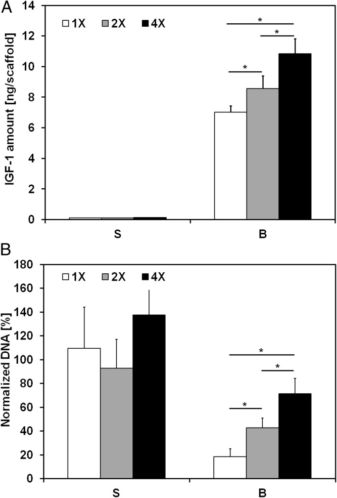
Fig. 5.
Effect of flow-derived shear stress on IGF-1R blockade within ES cells. IGF1 secretion and drug sensitivity for the experimental groups tested with different medium viscosities (S, static; B, bioreactor, with 1×: 0.9 ± 0.1 cP, 2×: 1.7 ± 0.1 cP, and 4×: 3.6 ± 0.3 cP) and a constant flow rate of 0.2 mL/min. (A) Amount of IGF1 ligand per scaffold measured in conditioned media after 10 d of culture. Error bars represent the SD (n = 3, *P < 0.05). (B) Cell viability after 3 d of culture followed by 7 d of exposure to dalotuzumab. Within each group, cell viability is shown as the DNA content of the drug-treated group normalized to the DNA content of the untreated group. Error bars represent the SD (n = 6, *P < 0.05).

