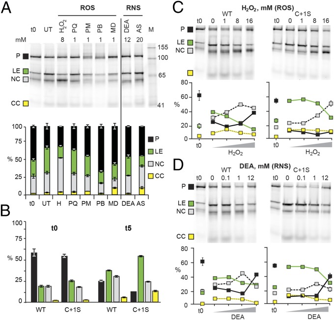
Fig. 3.
ROS and RNS modulate SufB splicing. (A) MIG SufB is sensitive to ROS and RNS. ROS (left of line) and RNS (right of line) treatments of MIG SufB at 5 h (t5). The stack plot shows the percentage of GFP-containing species. ROS compounds: H2O2 (H), paraquat (PQ), phenazine methosulfate (PM), plumbagin (PB), and menadione (MD); RNS compounds: DEA NONOate (DEA) and Angeli’s salt (AS). (B) C+1S MIG SufB mutant splices more readily than WT. The bar graph is derived from 0- and 5-h untreated samples (0 mM) of WT and C+1S MIG SufB from C. (C and D) H2O2 and DEA modulate splicing in a concentration-dependent manner. The gels are representative images of treatments of WT and C+1S MIG SufB, quantitated in scatter plots. Results with full-length exteins are similar (Fig. S3).

