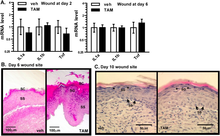
Fig. S3.
Gene expression and histology of healing wounds in young K14S mice. (A) mRNA levels of inflammatory cytokines, determined by qPCR, in wound areas of 8-mo-old veh- and TAM-treated K14S mice, at 2 d (veh and TAM, n = 3) and 6 d (veh, n = 5; TAM, n = 7) after injury. Mean ± SEM values with asterisks indicate differences at P < 0.05 by two-way ANOVA followed by Bonferroni post hoc analysis. (B and C) Representative photomicrographs of H&E staining of the wound area of 8-mo-old veh- and TAM-treated K14S mice at 6 d (B) and 10 d (C) after injury (veh and TAM, n = 5). Stratum basale (SB, arrows) are identified by strong nuclear staining (blue) at the bottom of the wound site. Stratum granulosum (SG, asterisks) are cells with granules in the cytoplasm. Stratum spinosum (SS) are cells with low nuclear staining above the SB layer. Stratum corneum (SC) is the outermost layer.

