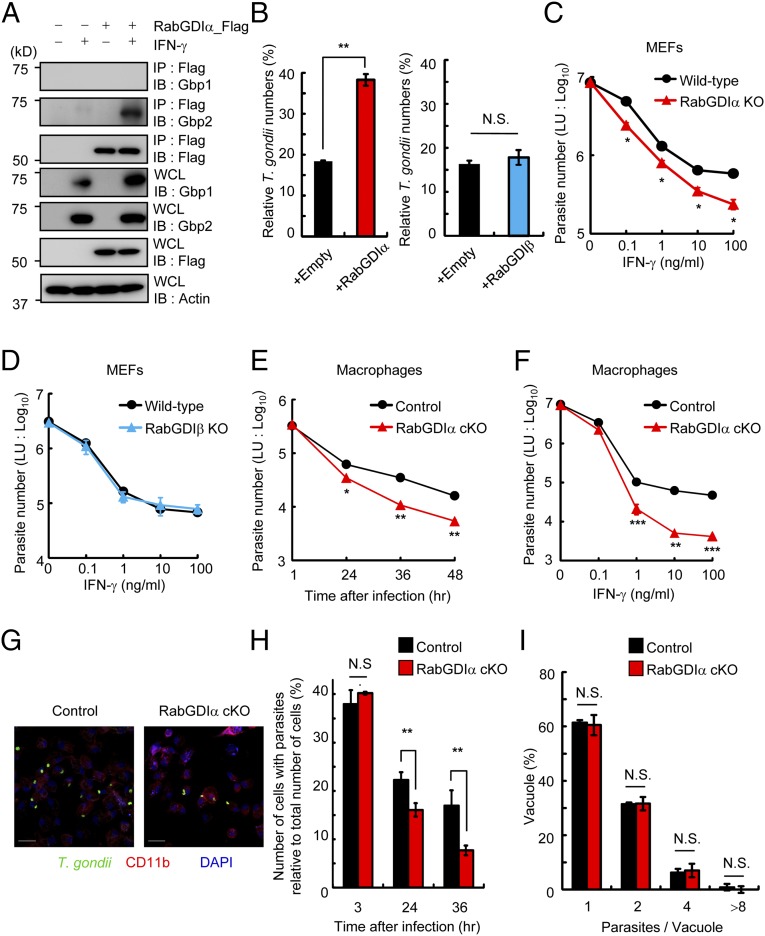
Fig. 1.
Enhanced IFN-γ–dependent T. gondii clearance in RabGDIα-deficient cells. (A) MEFs stably transfected with empty or Flag-tagged RabGDIα expression plasmids were untreated or treated with IFN-γ and lysed. The lysates were immunoprecipitated with anti-Flag and detected with the indicated Abs by Western blot. (B) T. gondii numbers at 36 h postinfection in control and MEFs overexpressing RabGDIα (Left) or RabGDIβ (Right) untreated or treated with IFN-γ were analyzed by luciferase assay. Percentages of parasite numbers (calculated by luciferase counts) in IFN-γ–stimulated control or cells overexpressing RabGDIα or RabGDIβ relative to those in the unstimulated respective cells are shown as “Relative T. gondii numbers.” (C and D) MEFs lacking RabGDIα (C) or RabGDIβ (D), and WT cells were untreated or treated with the indicated concentrations of IFN-γ. Untreated or IFN-γ–treated cells were infected with ME49 T. gondii expressing luciferase [multiplicity of infection (moi) = 1] and harvested at 36 h postinfection. The luciferase units (LUs) were assayed with the lysates. Error bars represent means ± SD of triplicates. (E) Control and RabGDIα conditional KO (cKO) macrophages stimulated with 10 ng/mL IFN-γ were infected with ME49 T. gondii expressing luciferase (moi = 0.5) and harvested at the indicated points postinfection. The luciferase units (LU) were assayed with the lysates. Indicated values are means ± SD of triplicates. (F) Control and RabGDIα cKO macrophages were untreated or treated with the indicated concentrations of IFN-γ. Untreated or IFN-γ–treated cells were infected with ME49 T. gondii expressing luciferase (moi = 0.5) and harvested at 36 h postinfection. The LUs were assayed with the lysates. Indicated values are means ± SD of triplicates. (G) IFN-γ–stimulated control and RabGDIα cKO peritoneal macrophages were infected with ME49 T. gondii (moi = 0.5), fixed at 36 h postinfection, and stained with rabbit anti-T. gondii (green) or rat anti-CD11b (red). (Scale bars: 20 μm.) (H) The percentage (number of cells with parasites relative to total number of cells) of control and RabGDIα cKO macrophages containing at least one parasite at the indicated points postinfection. Indicated values are means ± SD of triplicates. (I) The number of T. gondii parasites per vacuole in control or RabGDIα cKO macrophages at 36 h postinfection. Indicated values are means ± SD of triplicates. N.S., not significant; *P < 0.05, **P < 0.01, ***P < 0.001. Data are representative of three (A and C–F) and two (B and G) independent experiments. Data in H and I are pooled from two independent experiments in which almost 200 cells and 100 vacuoles were counted, respectively.

