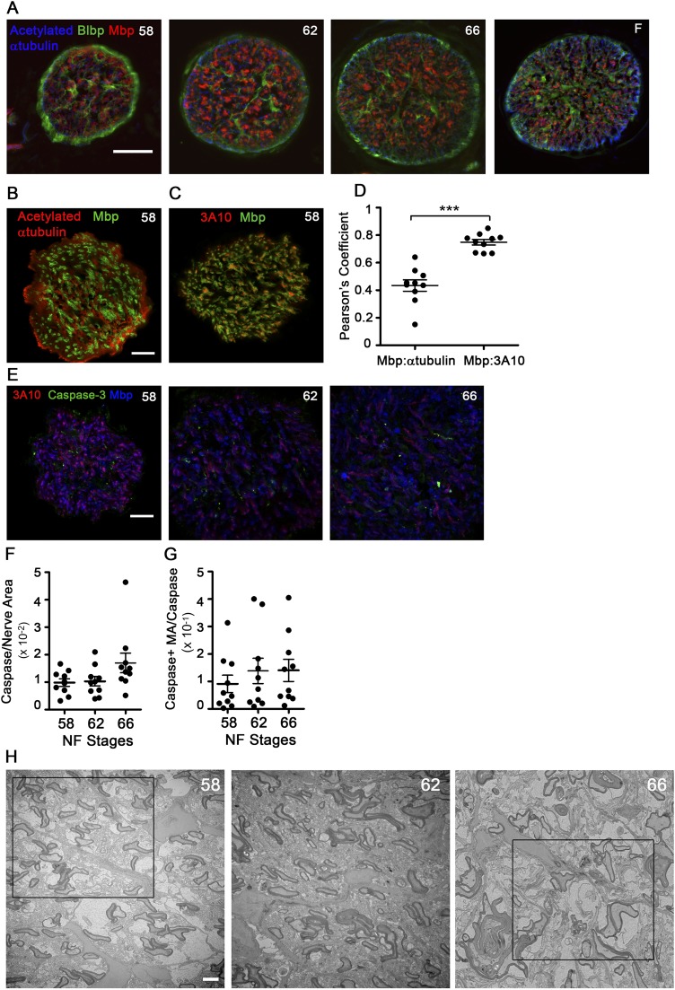
Fig. S1.
ON metamorphic remodeling does not lead to degeneration of myelinated axons. (A) ON cross-sectional cryosections immunolabeled for acetylated tubulin (blue, axons), Blbp (green, astrocytes), and Mbp (red, oligodendrocyte myelin). (Scale bar: 50 μm.) (B) Confocal images of stage 58 ON immunolabeled cross-sectional cryosections. All retinal ganglion cell (RGC) axons are labeled by acetylated tubulin (red), but only a subset of axons is surrounded by Mbp (green). (Scale bar: 20 μm.) (C) Confocal image of a stage 58 cross-sectional cryosection showing that the 3A10 neurofilament marker (red) preferentially labels axons bound by Mbp (green). (Scale bar: 20 μm.) (D) Pearson’s coefficient of colocalization between axon and myelin markers, derived from confocal images (n = 10 ONs). ***P < 0.001 by Student’s t test with Welch’s correction. (E) Confocal images of stage 58, 62, and 66 ON immunolabeled cryosections. Immunolabeling of myelin sheath labeled by Mbp (blue), large-caliber axons labeled by 3A10 (red), and degenerating axons labeled by cleaved caspase-3 (green) show no increase in degenerating myelinated axons at metamorphosis. (Scale bar: 20 μm.) (F) Cleaved caspase signal per unit area of ON measured from confocal images (n = 10 per stage) does not change during metamorphosis. (G) Cleaved caspase signal in myelinated axons per unit area measured from confocal images (n = 10 per stage) does not change over metamorphosis. (H) Representative TEM micrographs of ON cross-sections before tracing at stages 58, 62, and 66. (Scale bar: 2 μm.) Boxed areas are the areas shown traced in Fig. 1E. All measurements show mean ± SD.

