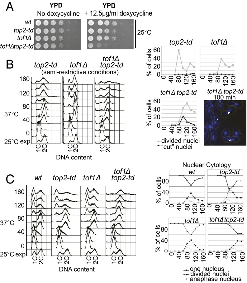
Fig. 4.
Deletion of TOF1 triggers excessive catenation of endogenous chromosomes and G2 cell-cycle arrest. (A) Viability assay of strains under the permissive condition of growth on YPD at 25 °C or with partial transcriptional repression of TOP2 (+doxycycline; DOX). (B) Deletion of TOF1 causes chromosome nondisjunction following partial depletion of Top2 activity. Cells were grown under semirestrictive conditions (YPD 37 °C + 12.5 µg/mL DOX) following synchronous release from G1 and analyzed for chromosome missegregation by FACS for DNA content (Left) or cytological analysis for cut and divided nuclei at the time points shown (min). Examples of cut cells (arrowheads) are shown (Right) [DAPI-stained DNA (blue) is shown over a light image of cells 100 min after release]. (C) The indicated strains were arrested in alpha factor, and Top2 was degraded (YP + 2% raffinose + 2% galactose; 37 °C + 25 µg/mL DOX). Samples were taken at midlog phase (25 °C exponential; exp) before alpha factor release (0) and at time points shown after release (min) for FACS analysis to assess DNA content (Left) or nuclear cytologies of cells (Right). The percentages of single (diamonds and solid line) and divided nuclei (squares and dashed line) are shown.

