Figure 2.
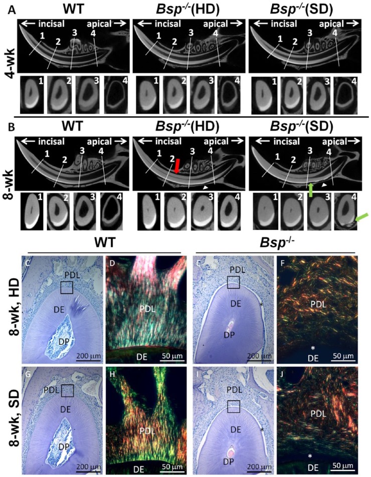
Altered incisor dentin and enamel formation in Bsp-/- mice. For micro–computed tomography (micro-CT) analysis, 4 landmarks were chosen: (1) alveolar crest, (2) mental foramen, (3) first molar, and (4) third molar. The wild-type (WT) images in panels A and B are representative of both hard diet (HD) and soft diet (SD) since no apparent differences in phenotype were observed between these groups. (A) At 4 wk of age, micro-CT analysis indicated no striking difference in incisors of Bsp-/- mice vs. WT. (B) By 8 wk of age, micro-CT images revealed increased dentin thickness (red arrow) and decreased pulp volume toward the apical end of the incisors in Bsp-/- mice as compared with WT, regardless of diet. In addition, resorption of enamel and dentin (green arrow) and incisor-associated bone (white arrowhead) can be seen in both Bsp-/- groups. (C, E, G, I) Histology (hematoxylin and eosin) confirmed increased dentin (DE) thickness and decreased dental pulp (DP) space in Bsp-/- mouse incisors vs. WT. (D, F, H, J) Detachment (*) and disorganization of periodontal ligament (PDL) collagen fiber bundles in Bsp-/- mouse incisors with Picrosirius red staining under polarized light. Soft diet does not normalize dentinogenesis, acellular cementum formation, or PDL attachment in Bsp-/- mouse incisors.

