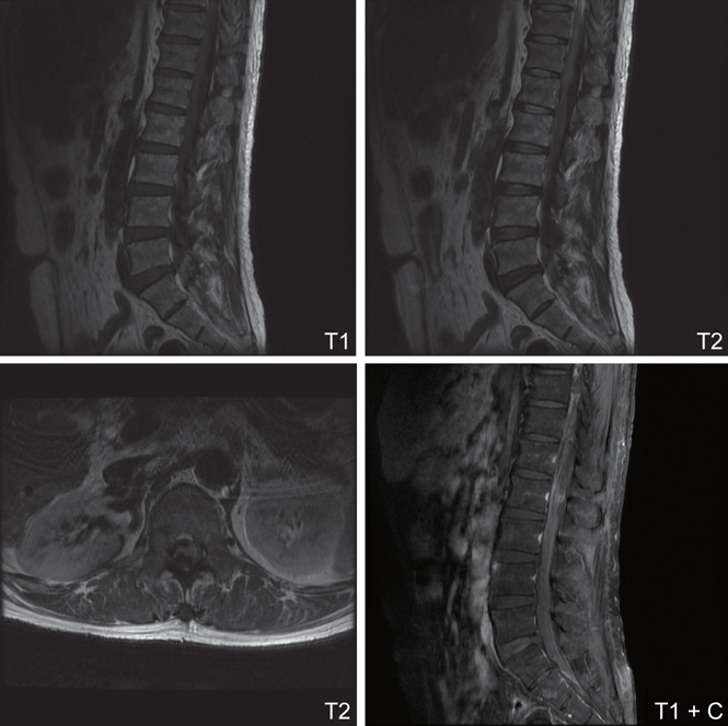
Fig. 1.

->Infiltrative hypointensity mass lesion in the dural sac from T12 to S1 level is noticed on both T1WI and T2WI. Acute hemorrhage was suspected. It compressed the spinal cord and cauda equinal nerves. Besides, there is an ill-defined intradural extramedullary mass lesion at L2 level, showing hyperintensity on T2WI and heterogeneous contrast enhancement on T1WI with contrast. Spinal metastatic tumor is highly suspected.
