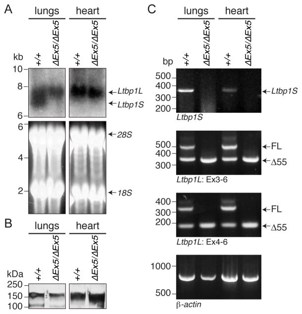
Figure 4. Ltbp1 ΔEx5 mice lack LTBP-1S but express Δ55 form of LTBP-1L.
(A) Northern blotting. Total RNA was extracted from newborn lungs and hearts of wild type and Ltbp1 ΔEx5 mice and utilized for Northern Blotting analysis. Ltbp1S transcripts were detected only in lungs of wild type samples, whereas transcripts of Ltbp1L were detected in both tissues of wild type and Ltbp1 ΔEx5 mice. (B) LTBP-1 protein analysis. Total protein extracts from newborn lungs and hearts of wild type and Ltbp1 ΔEx5 mice were subjected to plasmin digestion to liberate matrix-bound proteins. The released, truncated LTBP-1 was visualized by Western blotting with a rabbit polyclonal anti-LTBP-1 antibody (Ab 39). Bands around the predicted mass of 150-kDa were detected in all samples. (C) RT PCR. RT PCR was performed with lung and heart cDNAs from wild type and Ltbp1 ΔEx5 mice. The primer set specific for Ltbp1S amplified only wild type templates (top panel). Primers flanking exon 5 (Ex3-6 or Ex4-6) revealed expression of a full length (FL) and an Ltbp1L splice variant lacking exon 5 (Δ55) in wild type samples, whereas only Δ55 was amplified in Ltbp1 ΔEx5 samples (second and third panel, respectively).

