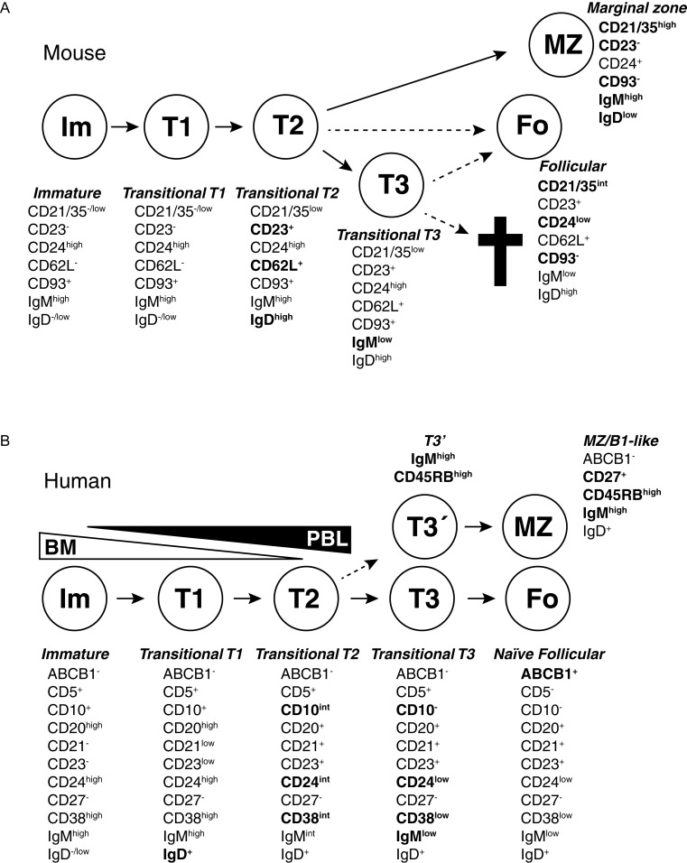
Fig. 2. Transitional B cells in mouse and humans.
B cells go through several stages after leaving the bone marrow but before becoming fully mature. During these stages, self-reactive cells are depleted due to interactions with self-antigens and are also selected into different lineages. The different stages can be defined based on expression of cell surface antigens. Changes important for defining different stages have been marked in bold. A: Several stages that mouse transitional T cells go through after leaving the bone marrow have been defined based on cell surface markers and transfer experiments. T1 cells in spleen are similar to immature B cells in the bone marrow. CD23 and IgD are upregulated when the cells become T2 cells, and T1/T2 cells are also precursors to MZ B cells. A T3 stage has also been defined. T3 cells were first suggested to be a pre-stage to mature follicular cells, but later analysis revealed that this population may be enriched for autoreactive cells and may be targeted for deletion through apoptosis due to interactions with self antigens. Cell phenotypes adopted from Allman and Pillai, 2008[8] in which the relationship between different transitional and more mature stages is discussed. B: Also in humans several transitional stages have been defined, going from immature B cells leaving bone marrow to naïve follicular B cells and circulating MZ-like/B1-like lineage B cells. Although transitional cells are termed T1, T2 and T3 in both humans and mice, there is not necessarily a direct correlation between them. Many expression changes during human transitional development are continuous, making it hard to set clear gates between different populations. The T3 population can be divided into two (T3 and T3′) based on expression of the CD45RBMEM55glyco-epitope and IgM, and CD45RBMEM55/IgMhigh T3 cells appear to be precursors to innate MZ/B1-like B cells. Cells with T1, T2 or T3 phenotypes have been identified both in peripheral blood and in the bone marrow. It is unknown if this is due to circulation of cells or if some cells go through these developmental stages within the bone marrow before release. Cell phenotypes are adopted from Bemark et al., 2012[17].

