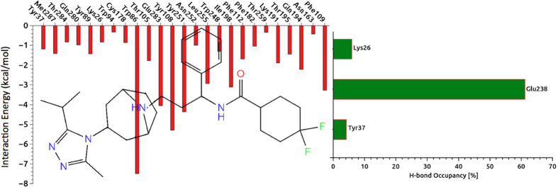
Figure 5.

(left) Non-covalent bond interactions between the amino acid residues at the binding pocket with the inhibitor. In order to identify individual amino acid residues participate in interaction with each segment of the ligand, the 2D chemical structure of maroviroc was inserted into the profile. (right) The occupancy in percentage of each residue participating in H-bonding with the inhibitor.
