Figure 7.
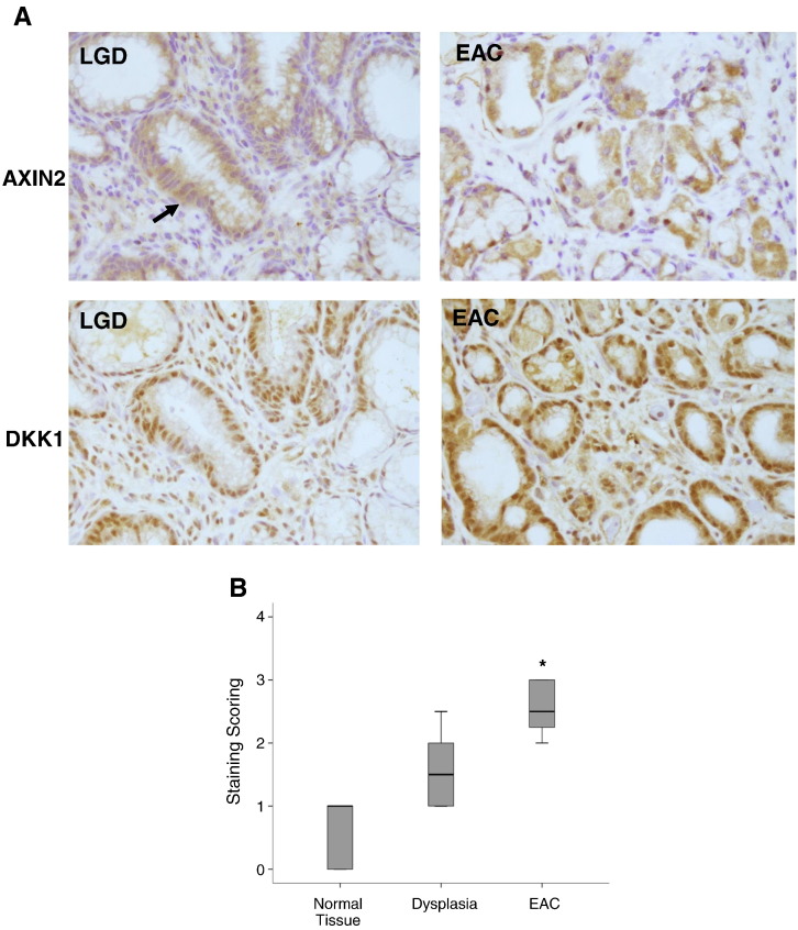
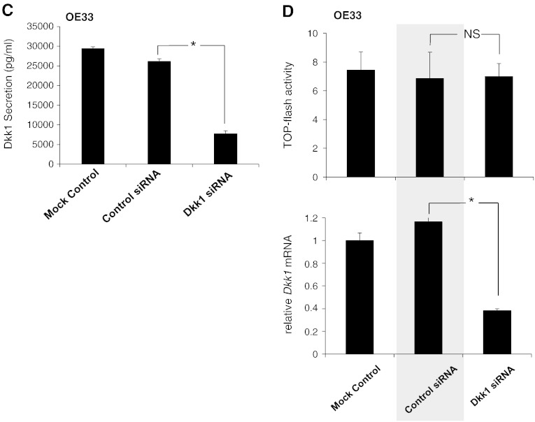
Dkk1 in BE-associated adenocarcinoma: (A) Representative immunohistochemistry for Axin2 and Dkk1 protein expression in esophageal tissues with BE-associated low-grade dysplasia (LGD) and EAC. The arrow indicates the area of the LGD, depicting large and irregular nuclei displaced from their normal position near the basement membrane. (B) Box plot analysis of immunohistochemical experiments for Dkk1. A simple grading system of 0 to 3 was used. Overall staining was evaluated in SQ (n = 8), LGD (n = 5), and EAC (n = 8). Median values are shown as thick lines (*P < .05; Mann-Whitney test). (C) ELISA of Dkk1 secretion in culture medium of OE33 cells following transfection with Dkk1-siRNA. Histograms depict the mean of three independent biological replicates (± SEM) (*P < .05 compared with control siRNA; t test). (D) Luciferase assay (top) and qRT-PCR analysis of Dkk1 gene expression (bottom) in OE33 cells following siRNA-mediated Dkk1 gene silencing. NS: nonsignificant (*P < .05; t test).


