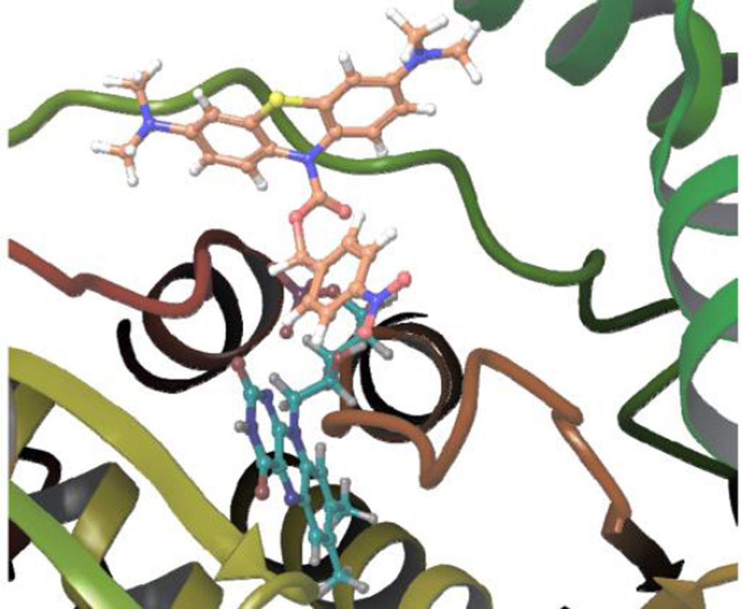
Fig. 5.
The best binding pose of p-NBMB (orange carbons) to NTR. FMN is shown as cyan carbons. The substrate binding pocket of NTR (PDB 1F5V) was generated using Fred v3.0.0 of OpenEye software suite. The enzyme is represented as cartoon and colored by secondary structure (prepared with Maestro14).

