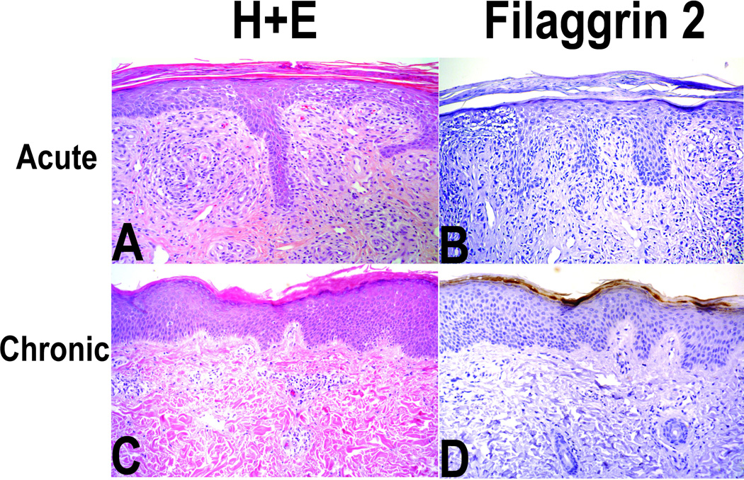
Figure 2. Typical histology (H & E and FLG2 immunostaining) of an individual with acute (panels A and B) and an individual chronic dermatitis (panels C and D).
Panel A (H & E) magnification 100X; acute spongiotic findings with grade 3 inflammation; Panel B (FLG2 staining) grade 1 FLG2 staining; Panel C chronic dermatitis with grade 1 inflammation; Panel D (FLG2 staining) grade 3/4 staining. Grading of FLG2 staining was based on the intensity of immunostaining using standard DAB technique.

