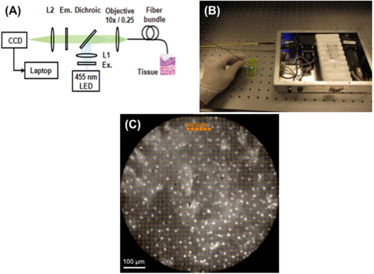
Figure 1.
(A) Device configuration. (B) The device is battery operated and easily fits in a briefcase. (C) To facilitate objective, real-time assessment of nuclear size and spacing, a grid with 19.4 μm spacing was superimposed on the display monitor and 15.1 μm diameter dots were placed; this image is normal esophageal mucosa.

