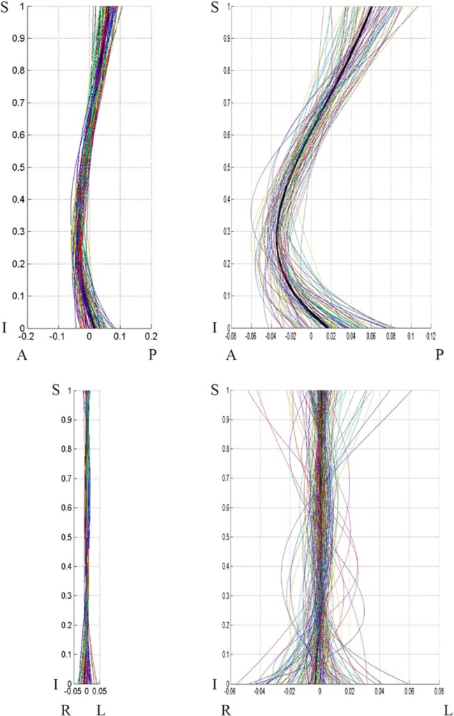
Fig 8. Female population curves, 2-D view.

Female population curve 2-D view: sagittal (top) and coronal (bottom) planes. The graphs on the left are scaled to the axes proportions; the graphs on the right are freely scaled to arbitrary proportions for a better view of the curve shape. Each line represents a curve sample. The coordinates are scaled by the vertical distance between the superior end plate of the sacrum and the inferior end plate of T12.
