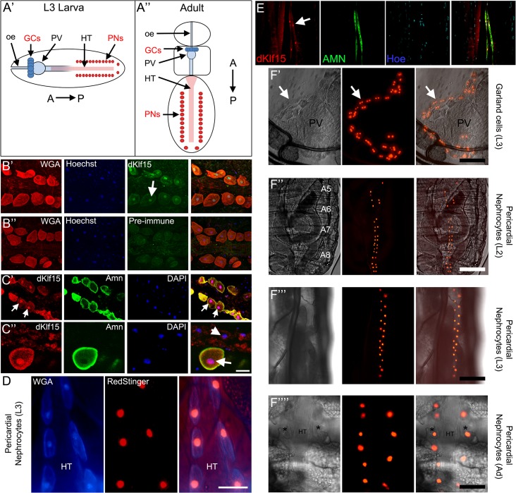
Fig 1. dKlf15 expression is restricted to nephrocytes.
(A’ & A”) Schematics showing location of the two nephrocyte populations in larval and adult Drosophila, the garland cells (GCs) and pericardial nephrocytes (PNs). Garland cells are at the interface between the paraventriculus (PV) and oesophagus (oe), whereas pericardial nephrocyte are either side of the heart tube (HT); A>P, anterior-posterior. (B’ & B”) Antisera raised to dKlf15 (but not the pre-immune sera, B”), locate to the nucleus of adult pericardial nephrocytes (arrow). (C) Two magnifications of the adult heart immunostained with anti-dKlf15 (1:10) and anti-Amnionless (1:100) antibodies. Arrows denote pericardial nephrocytes, the arrowhead indicates a cardiomyocyte nucleus. Scale bar = 20 μm. (D) The heart in a dissected L3 larva expressing the RedStinger fluorescent reporter driven by dKlf15-Gal4 and stained with wheat germ agglutinin which is taken up preferentially by pericardial nephrocytes (blue); HT = heart tube; scale bar = 40 μm. (E) L2 larva stained with anti-Amnionless and anti-dKlf15 showing localisation of dKlf15 to the nucleus of Amnionless-positive pericardial cells (arrow). (F’-F”“) dKlf15-Gal4 driven RedStinger expression in living larvae and a dissected adult. Fluorescence is seen in binucleate garland cells next to the paraventriculus (PV) in L3 larvae (arrows). Fluorescence was also detected in pericardial nephrocytes (asterisks) either side of the heart tube (HT) at L2, L3 and adult (Ad) stages. Scale bars = 50 μm.

