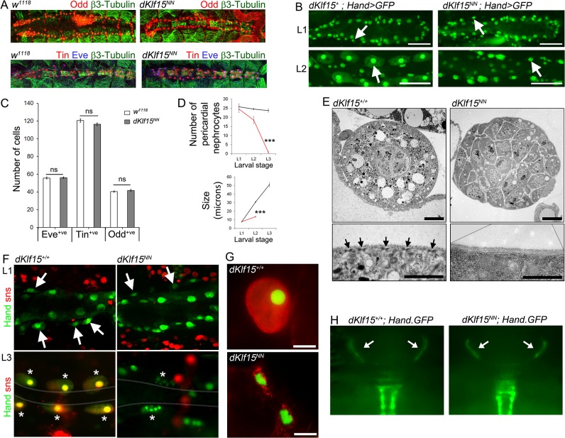
Fig 5. dKlf15 regulates the post-embryonic differentiation of pericardial nephrocytes.
(A) Stage 16 embryos stained with antibodies to Odd-skipped (Odd) or Even-skipped (Eve) and Tinman (Tin) and β3 tubulin. (B) Wild type (dKlf15 +) and dKlf15 NN on a Hand-GFP background to mark cardiomyocytes and pericardial nephrocytes (arrows). (C) Number of Even-skipped, Odd-skipped and Tinman positive cells in stage 16 embryos; n = 8–12 embryos per genotype. (D) Number and size of nephrocytes in larvae at different stages. ***P<0.001; n = 8–14 larvae per genotype; (note, nephrocyte were too infrequent to quantify at L3 stage). (E) Ultrastructure of nephrocytes from L3 stage wild type (dKlf15 +/+) and mutant dKlf15 NN larvae. Arrows indicate slit diaphragms. Scale bars = 5 μm (upper panels); = 1 μm (lower panels). (F) Hand-GFP wild type and dKlf15 NN lines expressing a sticks and stones reporter (red). Nephrocytes in L1 (arrows); nephrocytes in L3 (asterisks); grey line defines the heart. (G) Nuclear morphology of Hand-GFP-positive cells co-stained with wheat germ agglutinin (red), scale bar = 25 μm. (H) Micrographs show the Hand-GFP fluorescence signal of the ‘wing hearts’ (arrows) seen through the cuticle of the scutellum of pupa.

