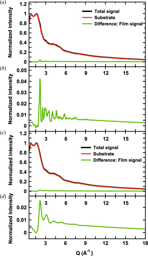
Figure 3.
(a) Normalized data collected for sample 1C (black) and a clean substrate (red). The difference curve is shown in green and is plotted on an expanded scale in (b). (c) Normalized data collected for sample 1A (black) and the clean substrate (red), and difference between the two (green), also shown on an expanded scale in (d).

