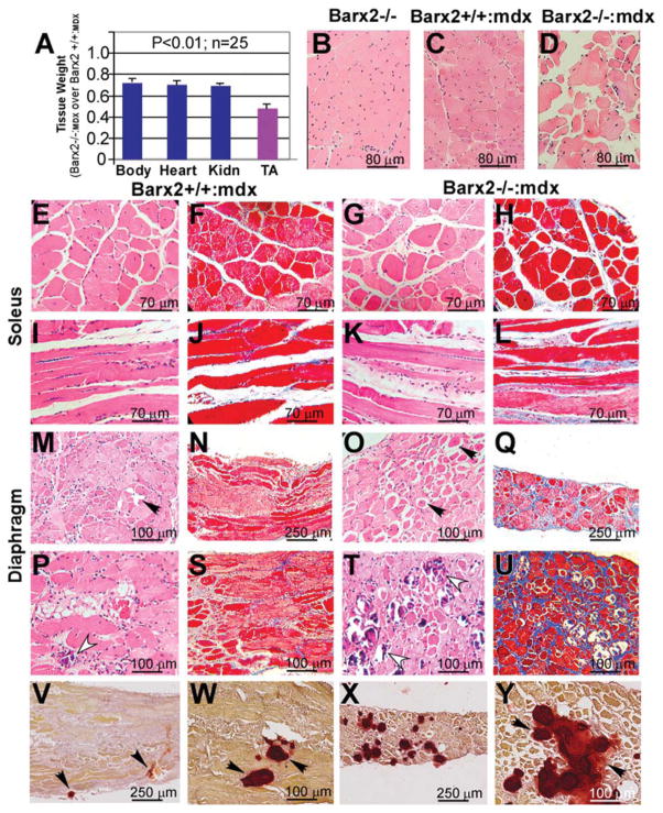
Figure 7.
Phenotype of Barx2/mdx double null mice. (A): Comparison of total body, heart, Kidn, and TA weights in Barx2−/−:mdx (shown as a ratio of Barx2−/−:mdx over Barx2+/+:mdx weights). TA muscle shows a dramatic reduction in weight relative to other organs. Tissues were harvested and weighed from 4-week-old Barx2+/+:mdx and Barx2−/−:mdx sibling pairs (n, number of pairs studied). (B–D): Hematoxylin-eosin (H&E)-stained paraffin sections of TA muscles obtained from 6-month-old Barx2−/−, Barx2+/+:mdx, and Barx2−/−:mdx mice. Double knockout TA muscle shows variability of fiber size suggesting presence of atrophy and hypertrophy of myofibers (D). (E–L): Soleus muscle of Barx2−/−:mdx mice (G, K, H&E staining; H, L, trichrome staining) shows greater variability in myofiber size and shape and marked endomysial and perimysial fibrosis relative to soleus muscle of Barx2+/+:mdx mice (E, I, H&E staining; F, J, trichrome staining). (M–U): Diaphragm muscles of 6-month-old (M–Q) and 12-month-old (P–U) Barx2+/+:mdx (M, P, H&E staining; N, S, trichrome staining) and Barx2−/−:mdx (O, T, H&E staining; Q, U, trichrome staining) mice. Diaphragms of 6-month-old Barx−/−:mdx mice appear much thinner than diaphragms of Barx2+/+:mdx mice (compare N and Q). In addition, diaphragms of 6-month-old Barx2−/−:mdx mice show frequent rounded atrophic fibers (black arrows). Atrophic fibers are dark red and glassy (they represent hypercontracted fibers). Trichrome staining shows more extensive fibrosis in six Barx2−/−:mdx diaphragms relative to Barx2+/+:mdx diaphragms (compare N and Q). The diaphragms of 12-month-old Barx2−/−:mdx mice show extensive fibrosis and greater instance of focal myofiber fibrosis (compare S and U), and necrotic myofibers undergoing myophagocytosis (T, white arrowheads) relative to Barx2+/+:mdx mice (P, white arrowhead). (V–Y): Alizarin red staining reveals increased calcium deposits in the Barx2−/−:mdx diaphragm muscle (X, Y) relative to Barx2+/+:mdx muscle (V, W). Abbreviations: Kidn, kidney; TA, tibialis anterior.

