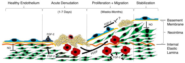
Figure 3.

Major molecular signals driving endothelial lining regeneration and neointima formation following denudation injury. Leftmost: Healthy endothelial lining suppresses smooth muscle proliferation through release of nitric oxide (NO). Middle left: Following denudation injury, damaged endothelial and smooth muscle cells release stored basic fibroblast growth factor (FGF-2). Platelets adhere to the site of denudation and degranulate to release stored platelet derived growth factor (PDGF). Monocytes adhere and enter the vessel wall, where they also secrete PDGF. Middle right: Following acute injury, PDGF stimulation results in smooth muscle cell migration from the arterial media toward the lumen, forming a neointima. FGF-2 stimulation results in the proliferation of both endothelial cells and smooth muscle cells. Endothelial proliferation is frequently incomplete. Rightmost: Depending on size and degree of medial damage, a functional endothelial lining may eventually regenerate and resume inhibition of smooth muscle proliferation via NO release.
