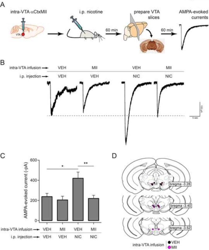
Figure 8. Inhibition of α6-containing nAChRs in VTA blocks AMPAR enhancement by systemic nicotine.

A) Experimental design. α6L9S mice were cannulated and vehicle or αCtxMII (10 pmol) was infused into the VTA. Following VTA infusion, mice were injected i.p. with saline or nicotine (0.03 mg/kg). Sixty min later, brain slices were prepared for recording. AMPA-evoked currents were elicited by locally puffing AMPA onto the cell body of the recorded neuron and recording inward cation currents in voltage clamp mode.
B) Representative AMPA-evoked currents from α6L9S mice injected/infused with the indicated drugs are shown.
C) Mean peak AMPA-evoked currents for each group shown in (B) are plotted. Mann-Whitney U-test: *p<0.05 (actual: p=0.0205), **p<0.01 (actual: p=0.003). (VEH/VEH: n=7, MII/VEH: n=7, VEH/NIC: n=8, MII/NIC: n=8)
D) Cannula location for each mouse in groups indicated in (B) is shown.
