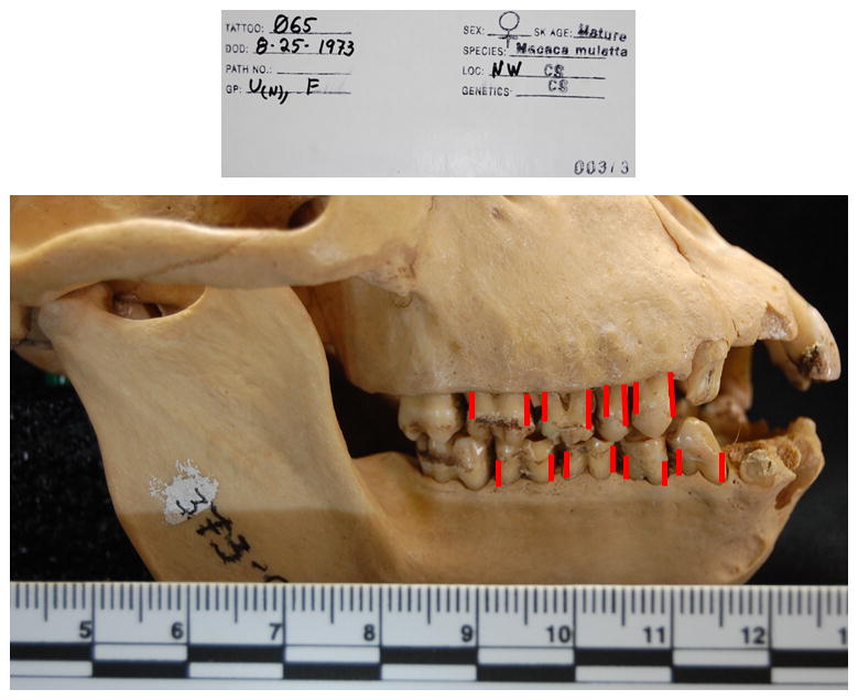
Figure 1.

Extraoral photograph of approach to evaluating periodontal disease in the skulls. Animal is 065, that died at 20.7 years of age in 1973. The red lines denote the measures obtained from the cementoenamel junction to the bone height on the distal and mesial buccal surfaces at mandibular and maxillary 1st and 2nd premolar and molar teeth. Measures were obtained from all four quadrants. The ruler is in centimeters.
