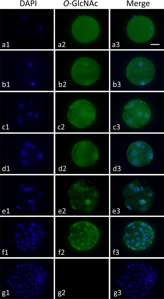
Fig. 2.
Localization of O-GlcNAcylated proteins in a pig metaphase II (MII) oocytes and parthenogenetically activated diploids during preimplantation development. MII oocytes (a1–a3) and diploids at the 2-cell (b1–b3), early 4-cell (c1–c3), late 4-cell (d1–d3), morula (e1–e3) and blastocyst stages (f1–f3) were treated with mouse anti-O-GlcNAcylated protein antibody (RL2) and then stained with anti-mouse IgG antibody labeled with Alexa Fluor 488 (green, center column). In the negative control (g1–g3), blastocysts were treated with only the secondary antibody. All diploids were counterstained with DAPI (blue, left column). Merged images are shown in the right column. Oocytes and diploids were observed under an epifluorescence microscope. Scale bar = 50 µm.

