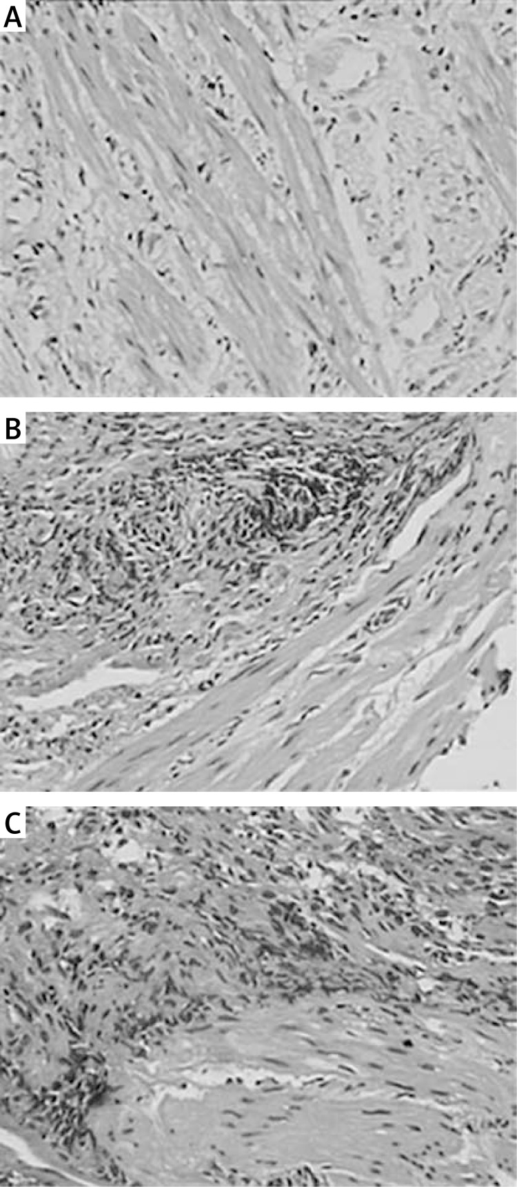
Figure 1.
H + E staining of collagen fibers. HEstained collagen fibers were observed under a light microscope (original magnification 200×). Normal collagen fibers were found in the control group, while, in the AS group, tissue hyperplasia of the collagen fibers and submucosal muscularis were observed. A – Control group; B – mild stenosis group; C – severe stenosis group

