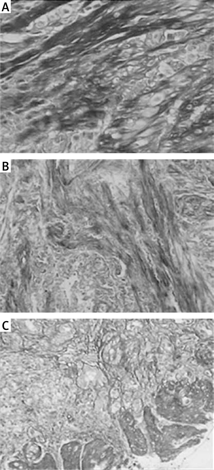
Figure 2.
Masson's trichrome staining of collagen fibers (original magnification 200×). The Masson's trichrome staining showed that distinct blue collagen fibers and red muscle fibers were observed. Compared with the control group, the deposition of reticular collagen fibers was more abundant in the mesenchyme of the AS group. The vessel wall became obviously thickened, and a narrow lumen was also observed in the AS group. A – Control group; B – mild stenosis group; C – severe stenosis group

