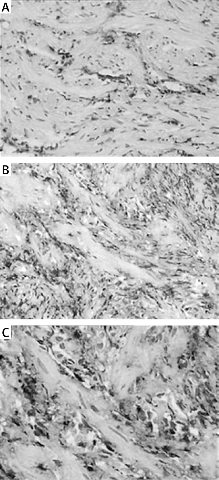
Figure 3.
Levels of TGF-β1 protein in the anastomotic tissues. Levels of TGF-β1 protein in tissue samples was determined by SABC IHC (original magnification 200×). Low expression of TGF-β1 protein was observed in the control group, while, in the AS group, levels of TGF-β1 protein were significantly increased, and the protein levels increased significantly (p < 0.01) with the increasing severity of AS. The positive expression of TGF-β1 protein in the AS group was indicated by the brown-yellow granules, mostly located in the cytoplasm of esophageal epithelial cells, endothelial cells, and fibroblasts. A – Control group; B – mild stenosis group; C – severe stenosis group

