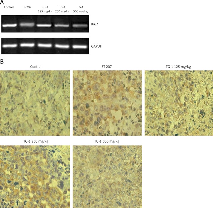
Figure 4.
RT-PCR detection of Ki67 mRNA and immunohistochemical staining for caspase-3 of the samples. A – Ki67 mRNA expression of transplant tumor OS-RC-2 treated with DMEM, FT-207, TG-1 (125 mg/kg, 250 mg/kg, or 500 mg/kg, respectively). Ki67 mRNA levels were measured by PT-PCR. GAPDH served as a control. B – Immunohistochemical staining for caspase-3 of the samples (400×). The positive reaction of the caspase-3 antigen showed brown particles in the nucleus and/or cytoplasm

