
Figure 1.
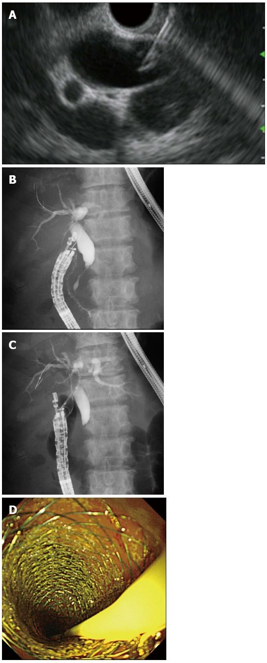
Endoscopic ultrasonography-guided choledochoduodenostomy. A: Endoscopic ultrasonography showing that extrahepatic bile duct was punctured by the needle; B: Guidewire was advanced through the needle into the right liver lobe; C: A self-expandable metallic stent (SEMS) was placed between the bile duct and the duodenum; D: Endoscopic view showing that the distal end of a SEMS was located at the duodenum.