
Figure 4.
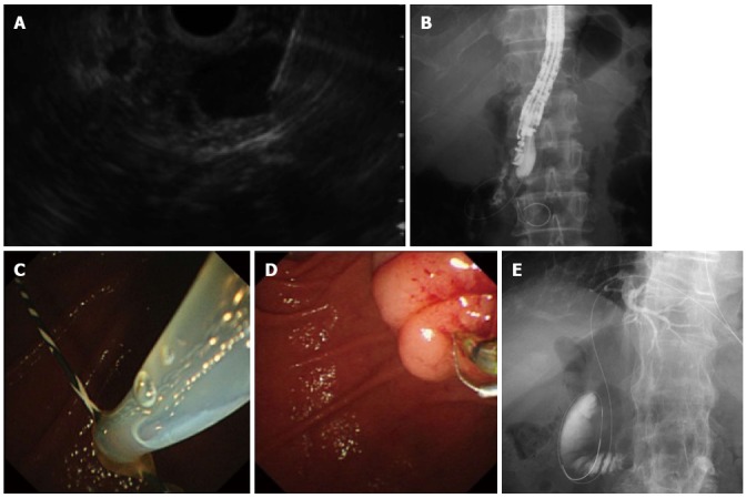
Endoscopic ultrasonography-guided rendezvous procedure. A: Endoscopic ultrasonography showing that extrahepatic bile duct was punctured by the needle; B: Radiography showing the guidewire was passed through the needle into the duodenum across the papilla antegradely. The endoscope was in the short position; C: Following echoendoscope withdrawal, the duodenoscopic view showed that a guidewire passing through the papilla was grasped by the snare; D: After the guidewire was pulled out through the accessary channel of the duodenoscope, a catheter was inserted into the bile duct over the existing guidewire; E: Transhepatic approach. Abdominal radiography shows that a guidewire was placed through the papilla into the duodenum.