
Figure 4.
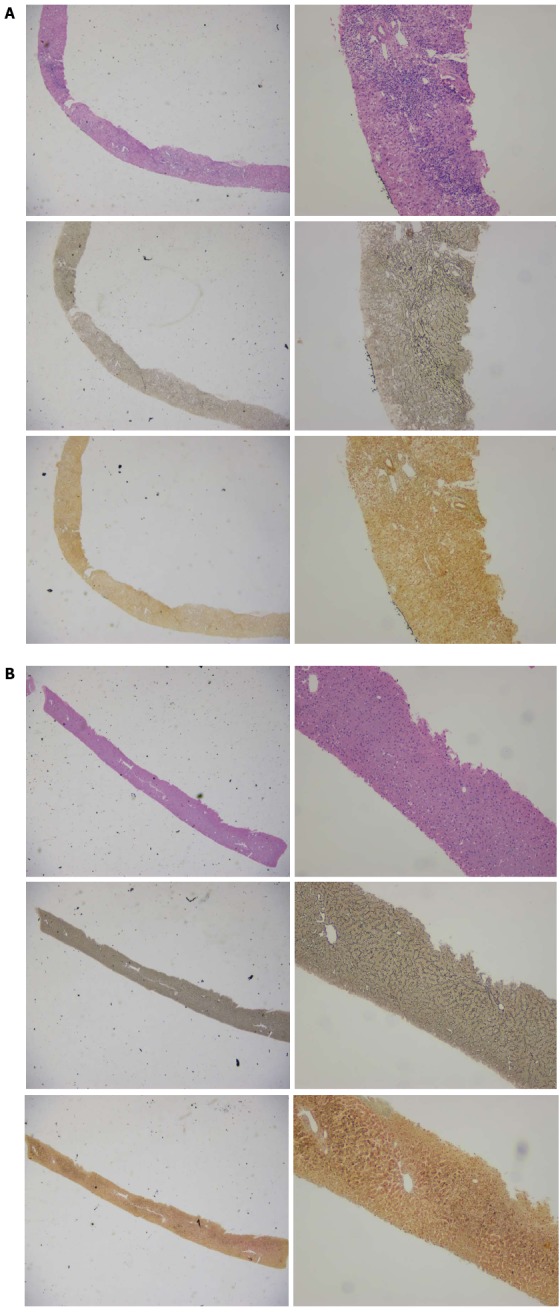
Pre- (A) and post-treatment (B) micrographs for a typical 35-year-old female patient with a favorable treatment outcome. Hematoxylin-eosin, reticular fiber and Masson’s trichrome staining was used in each row of the photos, respectively; × 20 for the left column and × 100 for the right column, respectively.