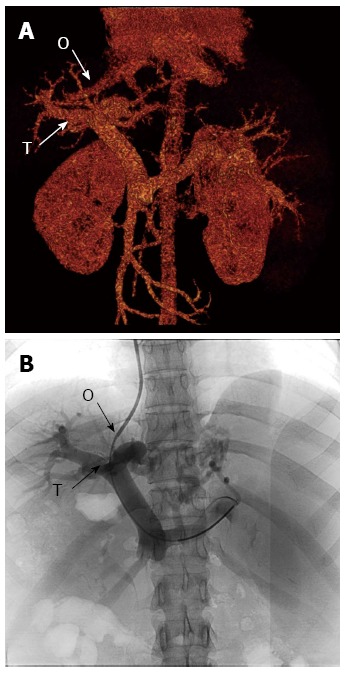
Figure 3.

Comparison of preoperative three-dimensional reconstructed image of the portal vein system and direct portogram obtained with non-iodinated contrast medium (Iopamiro 370) after a successful portal vein puncture. A: Preoperative three-dimensional reconstructed vascular image; B: Direct portogram of the portal vein (PV) during a successful puncture. O: Puncture point of the right hepatic vein; T: Target point of the right PV branch.
