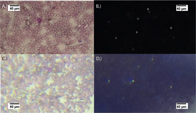
Figure 6.
Images of a Giemsa stained mouse blood smear using a Leica microscope with a 40X objective and (A) no-polarizers present in the image plane and (B) with a polarizer and analyzer crossed at 90 degrees in the image plane. The iPhone 5s utilized to acquire images of the same location on the prepared microscope slide with (C) the mobile phone polarized microscope system having no polarizers in the system and (D) the same system including a polarizer and analyzer crossed at 90 degrees in the image plane. In both crossed images, the birefringent hemozoin correspond well.

