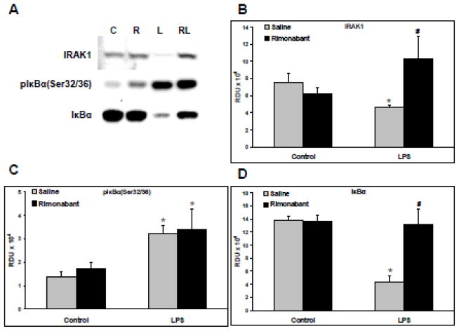
Figure 2. Rimonabant modulates TLR4 signaling following systemic LPS injection.
Following hemodynamic measurements, lungs isolated from rats exposed to systemic LPS (5mg/kg) for 30 minutes with or without intracerebroventricular rimonabant pretreatment (500 ng in 0.5 μl saline + 2.5% DSMO), were separated into left and right halves. Right lungs were homogenized, and supernatants, 20 μg/lane, were Western blotted. A) Representative blots are shown of IRAK1, phospho-IκBαSer 32/36, and IκBα; C=control, R=rimonabant, L=LPS, RL=rimonabant pretreatment + LPS. B) The Western blot band densities of IRAK1 in relative density units (RDU). C) The Western blot band densities of phospho-IκBαSer 32/36 in RDU. D) The Western blot band densities of IκBαSer 32/36 in RDU. Data are mean ± S.E.M. Significance was at P<0.05. * = significantly different from control. # = significantly different from its LPS-only group.

