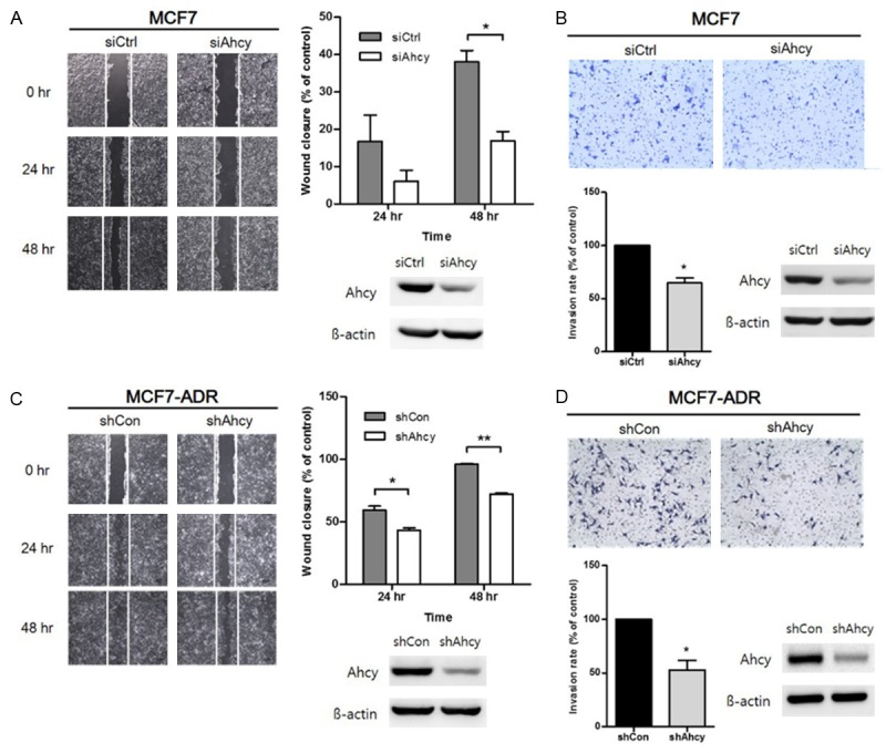
Figure 4.

Effect of inhibiting AHCY in breast cancer cells on cell invasion and migration. A, C. Effect of AHCY on cell migration was studied by wound-healing assay with both MCF7 and MCF7-ADR cell lines. A wound was made by scraping with a pipet tip and the plates were observed after 24 h and 48 h using microscope with 40 × magnification. The empty area was measured for quantification of wound closure rate using the Image J program. The expression of AHCY in these cells was confirmed by western blotting analysis. The graphs show mean ± SD of three independent experiments. *means P<0.05 and **means P<0.01. B, D. Invasive cells were observed after 24 h of seeding under a microscope with 100 × magnification. Quantification of the relative cell invasion was detected by 10% acetic acid solution. The expression of AHCY of cells used in each experiment was confirmed by western blotting analysis. The graphs show mean ± SD of three independent experiments. *means P<0.05.
