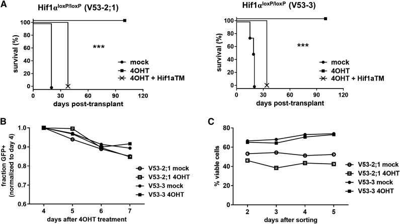
Figure 5.
Deletion of Hif1α eradicates LICs. (A) Survival of recipient mice after transplantation with leukemias deleted of Hif1α. Two independent mouse NOTCH1-ΔE leukemias on Hif1αloxP/loxP background were explanted, transduced with CreERT2/GFP only or CreERT2/GFP and Hif1αTM/Cherry viruses, and then treated with 4-OHT or vehicle (mock) control in vitro. GFP+ or GFP+ Cherry+ leukemia cells, respectively, were then FACS-sorted and injected into each of 4 recipient animals (n = 4) at a dose of 1 × 10⁵ cells per mouse. ***P < .001 (log-rank test). (B-C) Growth/survival of Hif1αΔ/Δ leukemia cells cultured in vitro. Primary mouse Hif1αloxP/loxP leukemias were explanted, transduced with CreERT2/GFP virus, and then treated with 4-OHT or vehicle (mock) in vitro. In (B), relative cell growth was assessed by tracking the % GFP+ cells over time, in culture by flow cytometry. A decreasing GFP+ fraction indicates transduced (GFP+) cells are growth disadvantaged compared with nontransduced (GFP−) cells in the same culture. In (C), transduced (GFP+) cells were FACS-sorted 2 days after initiation of 4-OHT treatment, then overall culture viability was assessed by flow cytometry for propidium iodide (PI) exclusion.

