
Figure 7.
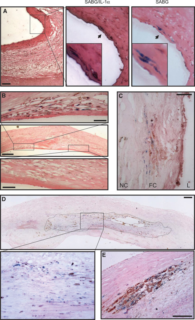
Senescent cells in human carotid plaques express interleukin-1α (IL-1α) and colocalize with IL-6 and CD68. Human carotid endarterectomy samples stained for senescence-associated β-galactosidase (SABG): showing senescent vascular smooth muscle cell (VSMC)-like cells in the fibrous cap that coexpress IL-1α (A); a discrete region containing many senescent cells showing localized IL-6 expression, in contrast to an adjacent region with no senescent cells or IL-6 (B); area within a fibrous cap (FC) showing high levels of local IL-6 expression adjacent to senescent cells (C); a large composite image showing localization between senescent and CD68 +ve cells (outlined and enlarged below; D); a region showing focal accumulation of senescent and CD68 +ve cells (E). Only eosin counterstain was used. Scale bars represent 100 and 25 mm (B; high power). L indicates lumen; and NC, necrotic core.