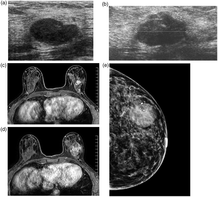
Fig. 1.
(a) A 40-year-old woman. US image at the time of initial core biopsy demonstrates a 1.8 cm well-circumscribed, hypoechoic lesion, consistent with a fibroadenoma. (b) Three years later, the FA is stable in size, but shows slight heterogeneity and is not as well circumscribed. (c) Fat-suppressed dynamic-enhanced MR image performed at the time of second ultrasound shows a 1.8 cm well-circumscribed, enhancing nodule. (d) Fat-suppressed dynamic-enhanced MR image 1 year after the previous MRI shows a heterogeneously enhancing mass that has doubled in size. (e) Left CC mammogram at the time of diagnosis of malignant phyllodes tumor shows a dominant mass with biopsy marker clip. Other marker clips are present in additional previously biopsied stable fibroadenomas.

