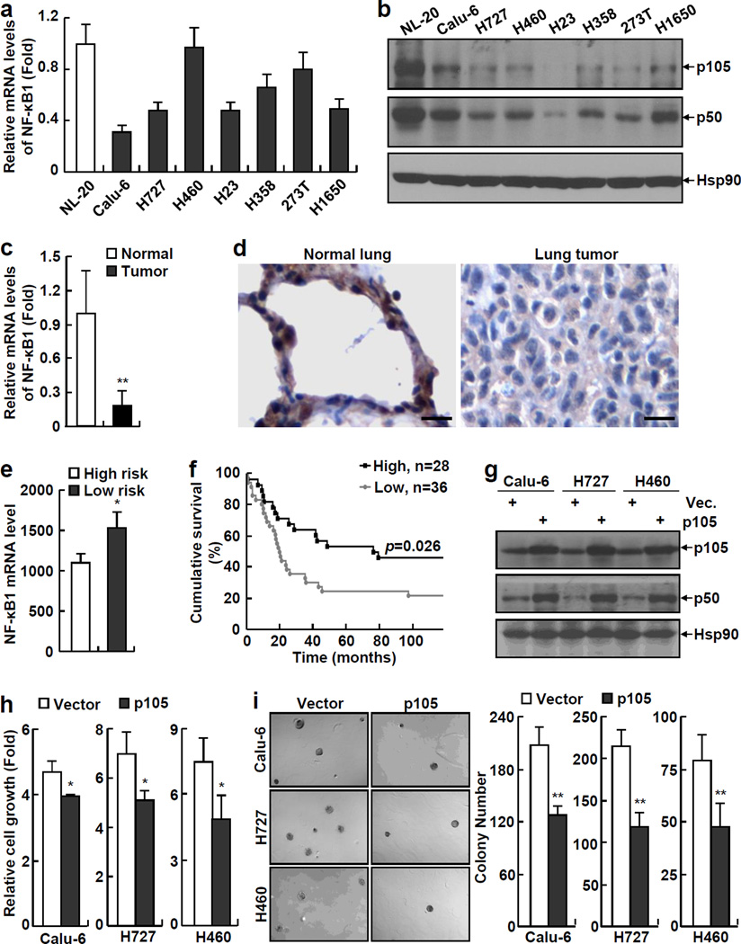
Figure 1.
NF-κB1 expression is decreased in human lung cancer cells and its re-expression inhibits the tumorigenicities of human lung cancer cells. (a) Real-time PCR assays showing decreased expression of NF-κB1 mRNA in human lung cancer cell lines. Normal human lung epithelial cell line NL-20 was used as a control. Data shown are means ± standard deviation (SD) (n = 3). (b) Immunoblotting (IB) assays showing decreased expression of NF-κB1 protein (p105 and p50) in human lung cancer cell lines. Hsp90 was used as a loading control. (c) Real-time PCR assays showing decreased expression of NF-κB1 mRNA in human primary lung tumor tissues. Normal control tissues from the same patients were used as controls. Data shown are means ± SD (n = 10; **, p < 0.01). (d) Immunohistochemical (IHC) analysis showing decreased expression of NF-κB1 protein in human primary lung tumor tissues. Scale bar: 20 µm. (e) Gene array assays showing an association between down-regulation of NF-κB1 mRNA and high risk of lung cancer in humans (The clinicopathological characteristics of patients used for the gene array were listed in the supplemental Table S1). (f) Gene array assays showing an association between low NF-κB1 mRNA expression and poor survival of patients with lung cancer. (g) IB assays confirming the expression of p105 and p50 proteins in NF-κB1 human lung cancer stable cell lines. (h) Cell growth assays showing decreased growth rate of NF-κB1 human lung cancer stable cell lines. Cells were cultured 3 days before growth assays. Data shown are means ± SD (n ≥ 3; *, p < 0.05). (i) Soft agar colony formation assays showing decreased anchorage-independent growth of NF-κB1 human lung cancer stable cell lines. Data shown are means ± SD (n ≥ 3; **, p < 0.01).

