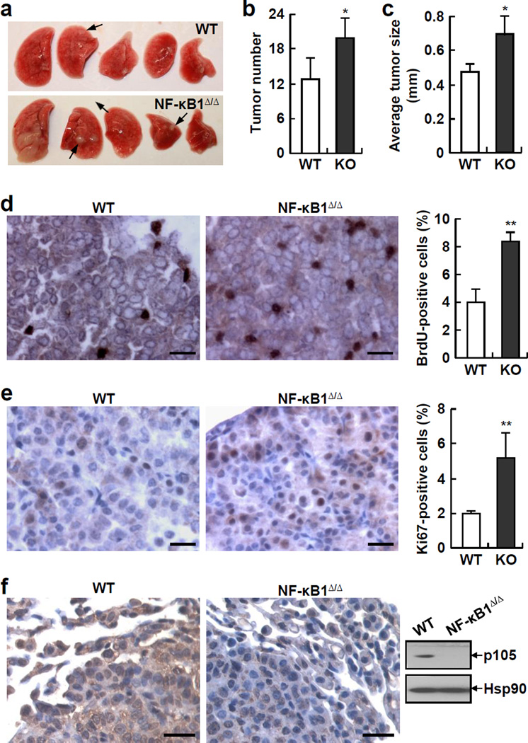
Figure 2.
NF-κB1 knockout (NF-κB1Δ/Δ) mice are more susceptible to urethane-induced lung tumorigenesis than wild type (WT) mice. (a) Lung tissues from urethane-treated NF-κB1Δ/Δ mice and WT mice. Representative tumors are indicated by arrows. The large white nodules in the most left lung tissue of NF-kB1Δ/Δ mice are not tumors but actually damages associated with increased inflammation (see Figure 3). (b) Increased lung tumor multiplicities in urethane-treated NF-κB1Δ/Δ mice. Data shown are means ± SD (n ≥ 6; *, p < 0.05). (c) Increased average size of lung tumors in urethane-treated NF-κB1Δ/Δ mice. Data shown are means ± SD (n ≥ 5; *, p < 0.05). (d) BrdU labeling showing increased proliferation rate of lung tumors in urethane-treated NF-κB1Δ/Δ mice. Scale bar: 20 µm. BrdU-positive cells were also counted and represented as the percentage of total cells. Data shown are means ± SD (n ≥ 5; **, p < 0.01). (e) Ki-67 IHC staining showing increased proliferation rate of lung tumors in urethane-treated NF-κB1Δ/Δ mice. Scale bar: 20 µm. Ki-67-positive cells were also counted and represented as the percentage of total cells. Data shown are means ± SD (n ≥ 5; **, p < 0.01). (f) IHC staining and IB assays showing absence of NF-κB1 proteins in lung tumors from urethane-treated NF-κB1Δ/Δ mice. Scale bar: 20 µm.

